EMF and the brain
Microwave Radiation and the Brain
Microwave Sickness and other symptoms from the brain
How does wireless effect the brain?
Wireless technologies can effect the brain by giving it stress and by oxidative stress mechanisms.
It can damage the brain and this can give various symptoms.
Stress. Stress symptoms are common symptoms.
Sleeping problems.
The blood-brain barrier can be damaged.
Cells can die.
Cancer cells like glioma can develop.
Behaviour and function can be effected like ADHD, autism ao.
Problems with concentrating
Speaking can be effected and language development can be slowed.
Mathematical thinking can be affected.
Nerves and myelin around the nerves can be damaged.
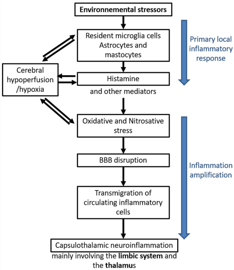
Inflammation in the brain.
Stress, irritability, and even aggression can be seen.
Microwave sickness is often shown by nerve pain in the brain.
Some get dizzy.
Often just some or a few of these symptoms are present.
Wireless technologies can effect the brain by giving it stress and by oxidative stress mechanisms.
It can damage the brain and this can give various symptoms.
Stress. Stress symptoms are common symptoms.
Sleeping problems.
The blood-brain barrier can be damaged.
Cells can die.
Cancer cells like glioma can develop.
Behaviour and function can be effected like ADHD, autism ao.
Problems with concentrating
Speaking can be effected and language development can be slowed.
Mathematical thinking can be affected.
Nerves and myelin around the nerves can be damaged.
Inflammation in the brain.
Stress, irritability, and even aggression can be seen.
Microwave sickness is often shown by nerve pain in the brain.
Some get dizzy.
Often just some or a few of these symptoms are present.

Microwave sickness info reflections and questions concerning the brain:
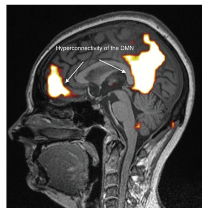
Heuser’s study where he brain scanned 10 people with EHS (microwave sickness) showed that all 10 people had hyperconnectivity in their medial orbitofrontal cortex.
The medial orbitofrontal cortex is associated with decision-making, emotional regulation, and reward processing.
Fire fighters who previously had been healthy developed several neurological problems after a mobile phone mast were put on the roof of the fire station. They got brain scanned just with another type of scan (the SPECT brain scan) and it showed damage all over the brain. (see links further down this page).
The study doesn’t explain the mechanisms behind.
Can this hyperconnectivity, where the region is more active than usual, be a compensatory mechanism or due to chronic stimulation?
Can it be the brain’s response to chronic pain or inflammation? Evidence suggests that chronic pain can alter brain connectivity, including increased activity in areas involved in pain processing and modulation.
It’s a point that Heuser explains how increased activity and increase in the brain (if it continuous) can lead to a decrease of the brain.
Other studies show that wireless technologies (like phones) can both stress but also damage the brain and leave to the death of some brain cells.
We’ve also noticed that BBB (blood barrier break) can occur.
Neurological effects can further be studied in The Bioinitiative report (link below).
Statistics of interest are:
beneign tumors in the forehead.
Malignant brain cancer (seen in the individual lopes).
Aggressive brain tumors like glioma types.
Stress
Neurological symptoms
Sleeping disorders
Behavioral problems
Learning disorders
Other symptoms.
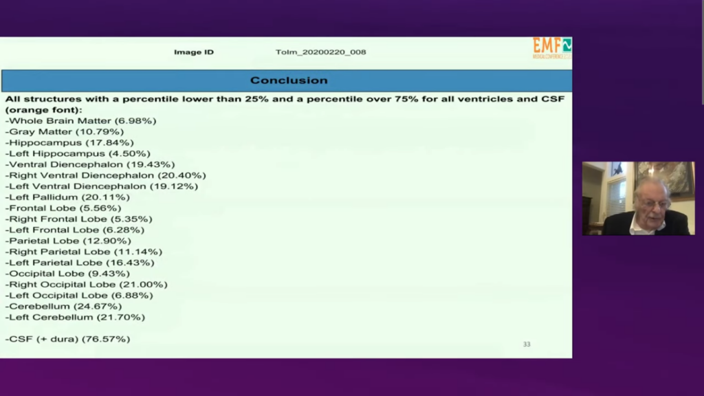
Functional brain MRI in patients complaining of electrohypersensitivity after long term exposure to electromagnetic fields
PubMed 2017
Gunnar Heuser , Sylvia A Heuser
The study made fMRI scans on 10 people suffering from EHS (microwave sickness).
“All ten patients had abnormal functional MRI brain scans. The abnormality was often described as hyper connectivity of the anterior component of the default mode in the medial orbitofrontal area. Other abnormalities were usually found. Regular MRI studies of the brain were mostly unremarkable in these patients.”
The study highlights hyperconnectivity in the anterior component (of the default mode network) in the area medial Orbitofrontal Cortex.
The medial orbitofrontal cortex is associated with decision-making, emotional regulation, and reward processing.
Link:
https://pubmed.ncbi.nlm.nih.gov/28678737/

The Critical Importance of Molecular Biomarkers and Imaging in the Study of Electrohypersensitivity.
2021 Jul 7
Belpomme among others.
A Scientific Consensus International Report.
International Journal of Molecular Sciences.
Pubmed
NIH National Institutes of Health
Link: https://www.ncbi.nlm.nih.gov/pmc/articles/PMC8304862/
PDF Test File
Effects of Radiofrequency Electromagnetic Radiation on Neurotransmitters in the Brain
EMR can lead to metabolic disorders and result in abnormal emotional behavior.
2021 Frontiers & PubMed
Cuicui Hu, Hongyan Zuo, Yang Li
Studies look at RF-EMR frequencies in the wifi and mobile phone radiation field.
- Exposure to electromagnetic fields can cause structural and functional changes in the nervous system.
- Many studies have shown that EMR affects the metabolism and transport of neurotransmitters. It is well understood that neural circuit is the structural basis of brain function, and the brain works by the interplay of various brain regions and many neurotransmitters. Consequently, the modulatory effect of EMR on neurotransmitter levels in various brain regions may play a critical role in the brain functioning.
- According to many studies, RF-EMR exposure can induce the imbalance of amino acid neurotransmitters in various parts of the brain.
- Several studies reported the effects of EMR on dopamine (DA).
- The results showed that the DA content in the brain tissue of fetal mice increased in the low-dose group but decreased in the high-dose group, and no significant changes were observed in the middle-dose group, which suggested that long-term mobile phone radiation could cause abnormal DA content in the central nervous system in fetal mice and might affect the brain development of mice.
- In summary, these studies indicate that EMR can lead to metabolic disorders of monoamine neurotransmitters in the brain, depending on the intensity of radiation exposure, and might in theory result in abnormal emotional behavior.
Links:
https://www.frontiersin.org/journals/public-health/articles/10.3389/fpubh.2021.691880/full
https://pubmed.ncbi.nlm.nih.gov/30562416/
Additional links;
https://nejtil5g.dk/aggression-og-hallucinationer-er-forbundet-med-tidlig-brug-af-smartphones/
https://nejtil5g.dk/jonathan-haidt-i-2012-faldt-de-unges-mentale-sundhed-ud-over-en-klippe/
https://www.emfdata.org/de/studien/detail?id=690
https://nejtil5g.dk/digital-uddannelse-en-vej-ud-af-uddannelseskatastrofen/
https://xn--die-pdagogische-wende-91b.de/aufruf-bildungspolitik-2025/
EMR may lead to abnormal norepinephrine (noradrenaline) and epinephrine (adrenalin) contents in the brain leading to neurotransmitter production disorders.
2021
EMR may lead to abnormal norepinephrine (noradrenaline) and epinephrine (adrenalin) contents (increase or decrease) in the brain leading to neurotransmitter production disorders.
- This further suggests that low-intensity EMR exposure can cause an increase in norepinephrine (=noradrenaline) content in the brain, which might in theory affect epinephrine (= adrenalin) content, leading to neurotransmitter production disorders.
- “The release of norepinephrine in the brain plays a role in various processes, such as stress, attention, sleep, inflammation, and the responses of the autonomic nervous system”.
- “these results suggest that long-term exposure to EMR may lead to abnormal norepinephrine and epinephrine contents in the brain, depending on the dose of radiation.”
Links:
https://www.frontiersin.org/journals/public-health/articles/10.3389/fpubh.2021.691880/full
EMR can lead to a decrease in excitatory amino acid neurotransmitters in the hippocampus, which may affect the excitatory-inhibitory balance of neurons, thus causing a decline in learning and memory ability
EMR exposure may reduce dopamine production in the hippocampus, affect rat arousal, and contribute to decreased learning and memory ability
2021 Frontiers & Pubmed
Cuicui Hu, Hongyan Zuo, Yang Li
“This study indicated that EMR exposure may reduce dopamine (DA) production in the hippocampus, affect rat arousal, and contribute to decreased learning and memory ability after exposure to EMR”.
- A decrease in DA was observed in the hippocampus of the EMR exposed group.
- there is a significant difference of DA and dihydroxyphenyl acetic acid (DOPAC) between hippocampus and striatum in the EMR exposed group
- a reduction in DA concentration in the striatum of mice.
- a certain intensity of microwave radiation can lead to abnormal metabolism of monoamine neurotransmitters in the hippocampus and striatum.
Links:
https://www.frontiersin.org/journals/public-health/articles/10.3389/fpubh.2021.691880/full
EMR induces brain injury: mood, feeding, cognition, memory, pain, sleep, and body temperature
EMR induces: decrease in learning and memory ability, abnormal hippocampal morphology and abnormal EEG results
2021 Frontiers & Pubmed
Cuicui Hu, Hongyan Zuo, Yang Li
- As an inhibitory neurotransmitter, 5-HT is mainly distributed in the pineal gland and hypothalamus, especially in the cerebral cortex and neural synapses. 5-HT contributes to the regulation of physiological functions such as mood, feeding, cognition, memory, pain, sleep, and body temperature maintenance, and these physiological functions have been reported as indicators of brain injury induced by electromagnetic radiation.
- These studies suggest that long-term exposure to microwave radiation can lead to an increase in 5-HT in the brain, indicating a disorder in the metabolism of the neurotransmitter.
- and these changes were related to the decrease in learning and memory ability, abnormal hippocampal morphology and abnormal EEG results induced by microwave radiation
Links:
https://www.frontiersin.org/journals/public-health/articles/10.3389/fpubh.2021.691880/full
EMR can reduce GABA neurotransmission, resulting in an imbalance in excitation and inhibition in the central nervous system.
EMR can cause metabolic disorders of the inhibitory neurotransmitters GABA and glycine, which may lead to neuronal dysfunction by affecting the neuronal excitation-inhibition balance.
2021 Frontiers & Pubmed
Cuicui Hu, Hongyan Zuo, Yang Li
Introduction:
“Metabolic studies have shown that all of the glucose is eventually converted to glutamate in the CNS, which indicating the key role of glutamate in multiple aspects of brain physiology.
Glutamate is the major excitatory neurotransmitter in the nervous system. Glutamate receptors distribute in neurons and glia of the brain and spinal cord.
In addition, glutamate also acts as a metabolic precursor to GABA and a component of various amino acid-based derivatives, such as the antioxidant glutathione.”
“GABA plays a critical role in the cerebral cortex, hippocampus, thalamus, basal ganglia and cerebellum, and has a regulatory role in various functions of the body, such as the regulation of emotion, memory and sleep, antihypertension, antifatigue, analgesia, etc. (63). GABA is produced in nerve endings catalyzed by glutamate decarboxylase. “
GABA is produced in the cytoplasm of presynaptic nerve endings. The enzyme glutamate decarboxylase (GAD) catalyzes the conversion of the amino acid glutamate into GABA.)
GABA, or gamma-aminobutyric acid, is the primary inhibitory neurotransmitter in the central nervous system, regulating neuronal excitability, and plays a role in other functions like sleep, stress, fear, and motor control. It works by reducing the ability of nerve cells to fire chemical messages, producing a calming effect that helps manage anxiety.
- The results showed that the contents of aspartate and glutamate decreased 1 day after radiation (10 min of microwave radiation), suggesting that acute EMR exposure could reduce the amount of excitatory amino acids in the hippocampus.
- The results showed EMR (1,800MHz EMR 1hour daily) induced significant decreases in glutamate and glutamine levels in hippocampal after 1 month.
- These data suggest that EMR can lead to a decrease in excitatory amino acid neurotransmitters in the hippocampus, which may affect the excitatory-inhibitory balance of neurons, thus causing a decline in learning and memory ability.
- Other data suggest the neurotransmitter disruption in the hippocampus might result in impairment of cognitive function caused by long-term microwave exposure.
- Wang et al suggests that decreases in NR2A, 2B and p-NR2B might contribute to the impairment of cognitive functions induced by microwave radiation (2.856 GHz, for 5 min/day, 5 days/week, over 6 weeks).
- The authors found that the levels of GABA and aspartic acid in the cortex and hippocampus significantly decreased after EMR exposure. These results suggest that EMR can reduce GABA neurotransmission.
- These results further suggest that microwave radiation may affect the neuroregulatory function of GABA, resulting in an imbalance in excitation and inhibition in the central nervous system.
- Overall, the above studies suggest that EMR can cause metabolic disorders of the inhibitory neurotransmitters GABA and glycine, which may lead to neuronal dysfunction by affecting the neuronal excitation-inhibition balance.
Links:
https://www.frontiersin.org/journals/public-health/articles/10.3389/fpubh.2021.691880/full
https://my.clevelandclinic.org/health/articles/22857-gamma-aminobutyric-acid-gaba
Effects of EMR acute microwave radiation exposure may result in cholinergic system dysfunction and a decrease in cognitive function
2021 Frontiers
Cuicui Hu, Hongyan Zuo, Yang Li
Introduction:
“The mode of action of Ach in learning and memory depends on the type of receptor it activates”.
“Cholinergic fiber projection from the basal forebrain to the cortex and hippocampus is the most important cholinergic system in the brain, and the cholinergic system plays a critical role in behavioral cognition. Ach is released from cholinergic nerve endings, and it was the first neurotransmitter to be measured in the brain. Changes in Ach in the extracellular fluid of the brain are closely related to functional changes in the central nervous system.”
- “This indicates that the increased synthesis and metabolism of Ach and the disordered expression of Ach receptors may result in cholinergic system dysfunction and a decrease in cognitive function in the early period of acute microwave radiation exposure.”
- “These studies further suggested that disorders of Ach synthesis and metabolism are an important part of the cognitive dysfunction caused by EMR.”
Links:
https://www.frontiersin.org/journals/public-health/articles/10.3389/fpubh.2021.691880/full
Ultrahigh frequency EMR resulted in a significant increase in Nitric Oxide (NO) may cause neuronal damage, which in turn leads to a decrease in learning and memory ability
2021 Frontiers
Cuicui Hu, Hongyan Zuo, Yang Li
- “Ultrahigh frequency EMR resulted in a significant increase in the level of Nitric Oxide (NO) synthesis in the mitochondria of neural cells in animal brain tissue and a significant increase in the activity of mitochondrial NO synthase. Considering the toxic effect of high NO concentrations on cells, the increase in NO may cause neuronal damage, which in turn leads to a decrease in learning and memory ability in mice.”
- Lai et al. reported that after 45 min of exposure to pulsed 2,450 MHz microwaves rats showed learning impairment. This indicated a deficit in spatial working memory function after EMR exposure.
- “This further suggests that both endogenous opioid neurotransmitter and cholinergic systems in the brain are involved in microwave-induced spatial memory deficits.”
- Mice were exposed to computer electromagnetic radiation. The results showed that the level of NO in the mouse brain gradually increased with prolonged radiation time.
- Ultrahigh frequency EMR resulted in a significant increase in the level of NO synthesis in the mitochondria of neural cells in animal brain tissue and a significant increase in the activity of mitochondrial NO synthase. Considering the toxic effect of high NO concentrations on cells, the increase in NO may cause neuronal damage, which in turn leads to a decrease in learning and memory ability in mice.
Links:
https://www.frontiersin.org/journals/public-health/articles/10.3389/fpubh.2021.691880/full

EMR induces an impairment of cognitive functions
https://www.frontiersin.org/journals/public-health/articles/10.3389/fpubh.2021.691880/full
https://pubmed.ncbi.nlm.nih.gov/30562416/
2021 Frontiers & PubMed
Cuicui Hu, Hongyan Zuo, Yang Li
“Numerous studies have shown that the nervous system is an important target organ system sensitive to EMR. Exposure to electromagnetic fields can cause structural and functional changes in the nervous system. Neurotransmitters are specific chemicals that act as messengers during synaptic transmission within the nervous system. Many studies have shown that EMR affects the metabolism and transport of neurotransmitters. It is well understood that neural circuit is the structural basis of brain function, and the brain works by the interplay of various brain regions and many neurotransmitters. Consequently, the modulatory effect of EMR on neurotransmitter levels in various brain regions may play a critical role in the brain functioning. According to many studies, RF-EMR exposure can induce the imbalance of amino acid neurotransmitters in various parts of the brain”
Links:
https://www.frontiersin.org/journals/public-health/articles/10.3389/fpubh.2021.691880/full
EMR exposure does increases the intracellular calcium and the formation of ROS, which would alter the cellular function eventually and lead to numerous biological effects including neurotransmitter imbalance.
The number of opened calcium channel increased with the presence of EMFs: The changes of intracellular calcium levels can trigger unusual synaptic action or cause neuronal apoptosis. This in turn can exert an influence on the neurotransmission of learning and memory process
Due to the dependent on oxidative phosphorylation for energy, neurons are vulnerable for oxidative stress compared to other cells. During EMR exposure, the occurrence of oxidant-antioxidant imbalance in the brain leads to oxidative stress
The various oxidants act to produce greatly elevated NF-kappa B activity, leading to inflammation and neural immune response, synaptic plasticity, learning and memory, neuroprotection and neurodegeneration.
2021 Frontiers
Cuicui Hu, Hongyan Zuo, Yang Li
“Some studies have suggested that the calcium activation could be the initial event leading to alteration in protein configuration, followed by generation of ROS and ultimately activation of the molecular apoptosis pathways.
Lushchak et al. reported that EMR exposure may firstly produce the free radicals in the brain and later they are converted to ROS. The elevation of ROS level can attack various biomolecules in the cell. The raised ROS can also in turn trigger calcium release, and then activate the genetic factors leading to DNA damage.
Any alteration in gene and enzyme levels, may result in the activation of downstream signaling, particularly the mitochondria-dependent caspase-3 pathway can cause the apoptosis of neurons, which would lead to altered behavioral manifestations and pathophysiological changes in the brain.
In a word, EMR exposure does increases the intracellular calcium and the formation of ROS, which would alter the cellular function eventually and lead to numerous biological effects including neurotransmitter imbalance. “
- “It is known that membrane is the first and an important target of EMF in cells. Cell membrane damage might result in neurotransmitter changes in the brain.”
- “EMR can alter cell membrane permeability such as changes in calcium, ionic distribution and ion permeability”.
- “Calcium is one of the important signaling substances, and an imbalance of calcium homeostasis can alter many functions of the cell. It was reported that the number of opened calcium channel increased with the presence of EMFs, which might resulting in the increased intracellular calcium concentration under EMR exposure. In addition, the changes of intracellular calcium levels can trigger unusual synaptic action or cause neuronal apoptosis. This in turn can exert an influence on the neurotransmission of learning and memory process”.
- It was reported that, EMR activation of voltage-gated calcium channels (VGCCs) causes a rapid increase in intracellular calcium, nitric oxide, and peroxynitrite.
- “The changes of membrane permeability may result in the damage of membrane integrity, and lead to the changes in brain neurotransmitter imbalance.”
- “It is known that neurotransmitter and its receptors are involved in various signaling related to cell proliferation, apoptosis, differentiation and inflammation. The crosstalk between neurotransmission and cell signaling may in turn affect the metabolism and transport of neurotransmitters. EMR exposures produce the main pathophysiological effects via excessive calcium signaling and the peroxynitrite pathway, and the diverse non-thermal effects of EMR are produced via VGCC activation”.
- “As the energy source of the cell, the mitochondrial calcium reaction was influenced by the alterations in calcium signaling pathways in response to the effects of EMR exposure”.
- “In addition to calcium signaling changes, EMR can cause activation of free radical processes and overproduction of reactive oxygen species (ROS) in neurons. Due to the dependent on oxidative phosphorylation for energy, neurons are vulnerable for oxidative stress compared to other cells. During EMR exposure, the occurrence of oxidant-antioxidant imbalance in the brain leads to oxidative stress”.
- ” The various oxidants act to produce greatly elevated NF-kappa B (NF-κB) activity, leading to inflammation. In addition, NF-κB signaling is reported to be involved in neural immune response, synaptic plasticity, learning and memory, neuroprotection and neurodegeneration.”
- “It has been shown that EMR exposure leads to up-regulated elements belonging to apoptotic pathways, which results in neuronal apoptosis. The probable mechanisms are mainly attributed to increased ROS generation following EMR exposure.”
- “The energy of non-ionizing radiation is not enough to directly break chemical bonds, and therefore the occurrence of DNA damage with non-ionizing EMR exposures is primarily a consequence of generation of ROS, followed by oxidative stress. Numerous animal experiments have clearly demonstrated that non-thermal EMR can cause oxidative stress, particularly in the brain”.
- “It has been documented that non-thermal EMR exposure of 900 MHz or 2.45 GHz in rats, either short-term or long-term, can trigger neuronal dysfunction and apoptosis of hippocampal pyramidal cells and cerebellum Purkinje cells through induction of oxidative stress.”
- “ROS plays a key role in the regulation of cytosolic calcium homeostasis. The protein phosphorylation and activation of the AP-1 family factors and nuclear factor kappa B (NF-κB) is regulated by the level of cytosolic calcium. Activation of the protein kinases pathways regulates the physiological response to EMR exposure including neurotransmitter imbalance, but the detailed mechanisms are still unclear.”
- “Many evidences indicate that EMR alter several aspects of calcium function in cells.”
- “Though we narrow down to biochemical imbalance to simplify explanation for changes of each neurotransmitter, the combined effects of neurotransmitters still deserves attention. It is also possible that the various neurotransmission effects following EMR exposure in animals might be due to combined effects in various brain regions, such as neurophysiological changes, increase of calcium and ROS, and thereby cell membrane damage and the downstream signaling changes. An imbalance in the excitation-inhibition imbalance of neurons resulting from neurotransmitter changes, would alter behavior”.
- “Consequently, the regulation of neural circuits may be involved in the neurotransmitter disorder of the brain induced by EMR.”
Links:
https://www.frontiersin.org/journals/public-health/articles/10.3389/fpubh.2021.691880/full
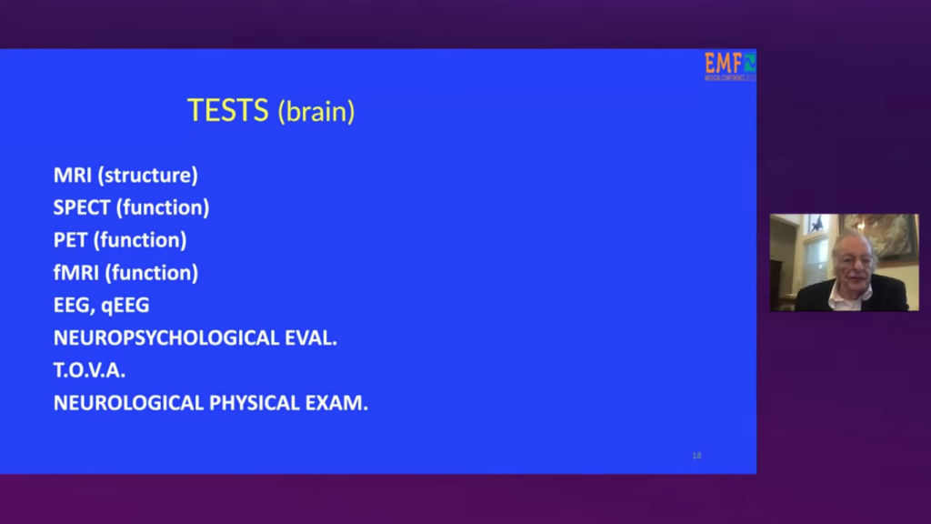
Neuroimaging methods
2021 Frontiers
Cuicui Hu, Hongyan Zuo, Yang Li
- “Several neuroimaging methods are used to illuminate the interference between brain electrical activity and EMR. For example, the changes of extracellular electrical potential in the cortex can be measured by EEG techniques, the regional changes of blood oxygen utilization can be detected with functional magnetic resonance imaging (fMRI) method during neuropsychological performance, and positron emission tomography (PET) reflects the cerebral metabolism”.
Links:
https://www.frontiersin.org/journals/public-health/articles/10.3389/fpubh.2021.691880/full
Additional information about GABA
Microwave Sickness Info writes:
GABA has caught our attention because study (see above) has shown that “EMR can reduce GABA neurotransmission, resulting in an imbalance in excitation and inhibition in the central nervous system.” from the study” Effects of Radiofrequency Electromagnetic Radiation on Neurotransmitters in the Brain”.
Here are some additional information about Gaba.
Did you know that GABA regulates stress and anxieity?
- GABA (gamma-aminobutyric acid) is the main inhibitory neurotransmitter in the adult central nervous system (CNS). Its primary role is to reduce neuronal excitability and regulate nerve cell activity.
- GABA is synthesized from the excitatory neurotransmitter glutamate.
- GABA dampens nerve impulses by binding to specific GABA receptors on the surface of neurons.
- Calming Effect: By reducing overall brain activity, GABA helps to regulate mood, decrease anxiety, stress, and fear, and promote relaxation and sleep.
- Anxiety and Stress: Low levels of GABA are linked to increased anxiety and stress responses.
- Epilepsy: Dysregulation of the GABA system can contribute to seizure activity.
- Sleep Disorders: GABA plays a vital role in regulating the sleep cycle, and deficiencies can lead to insomnia.
- Other Conditions: Reduced GABA function has also been implicated in conditions like ADHD, schizophrenia, autism, and Huntington’s disease.
https://www.frontiersin.org/journals/public-health/articles/10.3389/fpubh.2021.691880/fullhttps://pubmed.ncbi.nlm.nih.gov/30562416/
https://my.clevelandclinic.org/health/articles/22857-gamma-aminobutyric-acid-gaba

How does wireless radiation stress the brain?
You can measure on an EEG how wireless devices stress the brain.
Internal link to the test: https://microwavesicknessinfo.com/index.php/how-does-wireless-harm/
This video is based on the findings of neurobiology.

Wi-fi stresses the brain
Brain waves
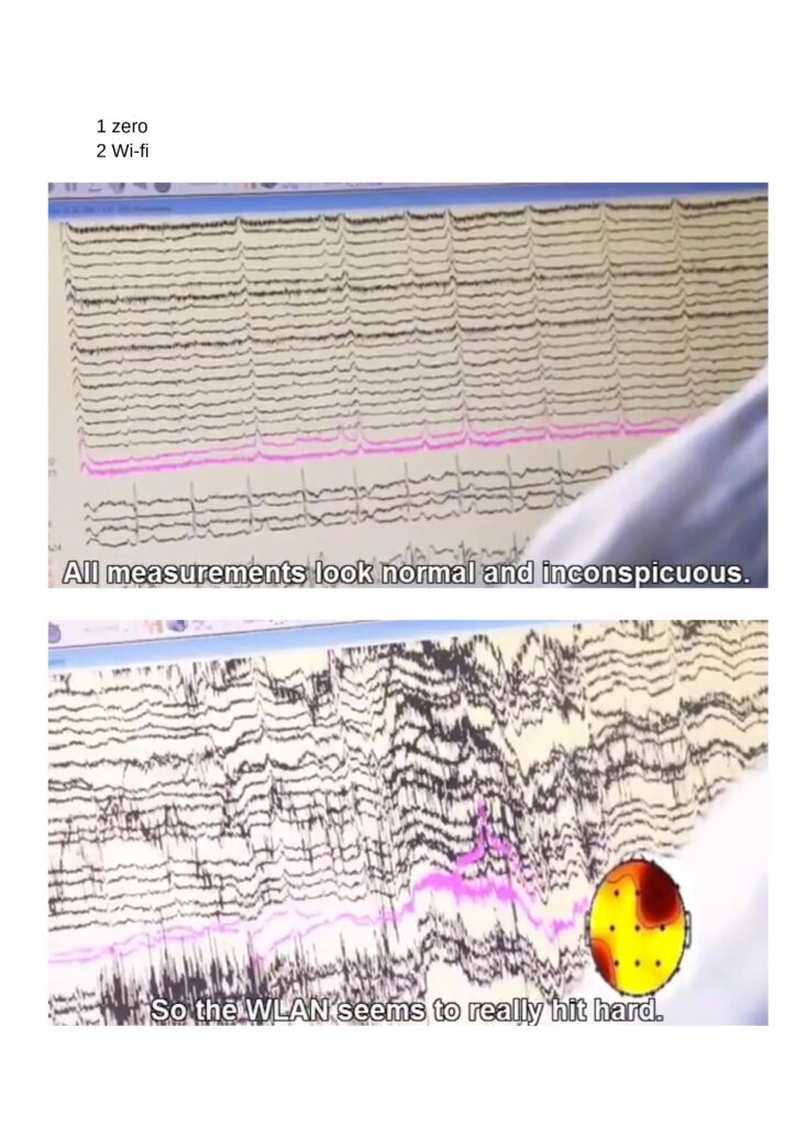
Here you can see how wi-fi stresses the brain.
Picture 1:
The first picture shows an EEG where the brain waves have been measured. The measurements are normal.
There’s no wi-fi. The wi-fi is turned off.
Picture 2:
Here you can see the brain waves measured with wi-fi (WLAN) turned on. The EEG measurement shows an abnormal level af stress on the brain.
You can watch the full video here on our page “How does wireless harm?”:
Growing up Healthy in a World of Digital Media
This video is based on the findings of neurobiology.
1. dec. 2021
What parents and educators should know:
Digital media affects brain development.
A video for parents, educators, teachers and parents’ evenings. Smartphones, tablet PCs, PC gaming – the world in which our children live is changing rapidly.
What do children need for healthy brain development?
What are the risks of smartphones and surfing the Internet?
How does cell phone and Internet addiction develop, what does sensory overload do to children?
This film shows how these risks can be avoided? We want our children to grow up to be healthy, self-confident and intelligent people who can later cope well with the complex demands of life.
For this to succeed, every child needs a variety of sensory experiences, it needs very different movements, motivation and empathy. This is how the child’s brain can develop in an all-round way.
This video is based on the findings of neurobiology.
Link to website:
https://shop.diagnose-funk.org/Buch-D…
Read about neurological effects in The Bioinitiative Report
https://bioinitiative.org/research-summaries/
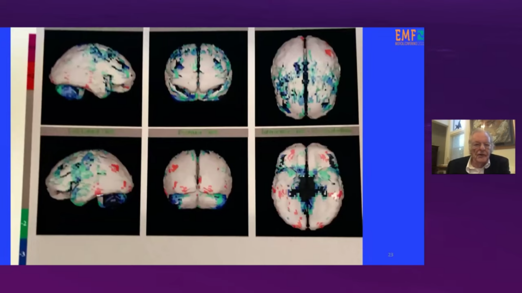
22 Functional Brain Scans of Patients Exposed to Neurotoxic Chemicals and/or EMF, Gunnar Heuser MD
This video is based on the findings of neurobiology.
fMRI brain scans
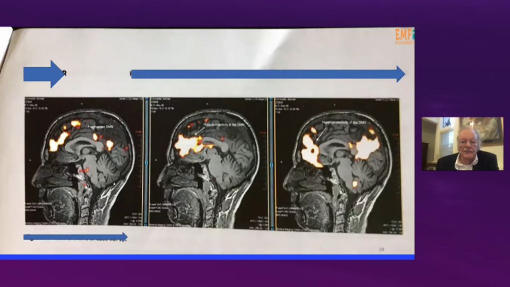
Gunnar Heuser MD explains following in the video.
fMRI brain scans
The brain scans show abnormality.
10 patiens with EHS symptoms were scanned and all brain scans showed abnormal results.
Scan showed connectivity and the brains were hyperconnected (can be seen on the images).
All 10 people had hyperconnectivity in the anterior component (of the default mode network) in the area medial orbitofrontal cortex.
Internal link to the test:
https://www.youtube.com/watch?v=s6M9nLG9Onw
SPECT brain scans
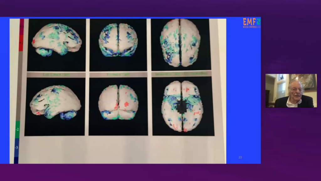
6 firefighters working under mobile phone mast at the fire station.
The firefighters were healthy before the mobile phone cell towers were put up.
The firefighters had problems with cognition, problems finding the way when driving, headaches, disorientation, and neurological problems.
MRI brain scans
Gunnar Heuser MD explains following in the video.
MRI brain scans
He explains how increased activity and increase in brain if it continuous can lead to a decrease of the brain (in the areas).
These data is from an MEI scan of an electrical lineman. He suffered from seizures.
Small amounts of repetitive stimulation can give seizures.
His right occipital lope is much larger and his left lope is much smaller.
Increasing Numbers of Children Aged 5-19 Years with Memory Problems in Sweden and Norway
2025
Mona Nilsson BSc and Lennart Hardell MD, PhD
“The data presented here show a very disturbing trend with a rapidly increasing number of children and adolescents seeking medical attention for memory problems or being diagnosed with mild cognitive impairment, especially over the past five to 14 years in Norway and Sweden. The increase cannot be explained by changed diagnostic criteria or reporting frequency.”
“What changes in children’s environment and lifestyle may have contributed to this worrying trend of impaired memory and cognitive function? There are several reports, both from animal studies and epidemiological studies in humans, that show that exposure to microwave RF radiation negatively affects memory and cognitive function. The hippocampus in the brain is important for short- and long-term memory, learning, and spatial navigation [41,42]. Several animal studies have also reported damage to the hippocampus from exposure to microwave RF radiation. Children’s exposure to microwave RF radiation from wireless technologies such as smartphones, base stations/antennas, Wi-Fi routers, smart meters, and other wireless technologies that emit microwave RF radiation has also increased rapidly during the same period as the increasing number of children with impaired memory and cognitive function.”
Increasing Numbers of Children Aged 5-19 Years with Memory Problems in Norway
Norway
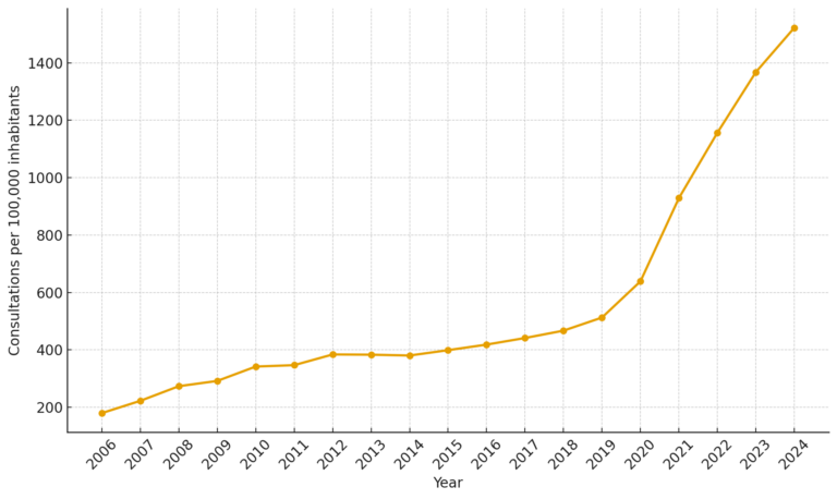
2025
Mona Nilsson BSc and Lennart Hardell MD, PhD
“Results
The data from Norway showed that there has been a clear
increase in number of consultations among children aged
5-19 years seeking medical care for memory problems each
year between 2006 and 2024″.
Links:
https://www.diagnose-funk.org/aktuelles/artikel-archiv/detail&newsid=2205
Increasing Numbers of Children Aged 5-19 Years with Memory Problems in Sweden
Sweden
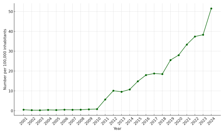
2025
Mona Nilsson BSc and Lennart Hardell MD, PhD
Results
“Data from Sweden shows rapidly increasing number of children aged 5-19 years diagnosed each year with R41.8
(mild cognitive impairment, subjective) that includes memory
problems.”
2008: iPhone smartphones were introduced in Sweden. They quickly became popular, as did other smartphones.
2009/2010: 4G is being rolled out.
2010: iPads and other tablets are introduced in Sweden. Wi-Fi and wireless computers or tablets are increasingly used in Swedish schools.
2016: Most schools had Wi-Fi. 4G+ is starting to be rolled out, increasing overall microwave RF radiation exposure over the next few years.
2017: Airpods and similar products are introduced in Sweden and are increasingly used in the following years among teenagers and children.
2019/2020: The rollout of 5G begins, causing a much higher overall exposure to microwave RF radiation in the following years.
2022: “The use of mobile phones by children and teenagers increased in Sweden. In 2022, more than 70% of Swedish children aged 15 used a smartphone for more than 3 hours a day. More than 40% of children aged 12 used it for more than 3 hours a day, “
Links:
https://internetmuseum.se/utstallningar/for-unga/nar-vara-mobiltelefoner-plotsligt-blev-smarta/
Increasing Numbers of Children Losing Competences in Germany
2025
Germany Children in schools losing competences from 2011-2021
“In light of years of digitalization initiatives in schools, the 75 experts draw a sobering conclusion: Academic performance in the core competencies of reading, writing, and arithmetic continues to decline, as does the overall level of education.”
““Digitalization in schools has not led to better educational outcomes – quite the opposite,” analyzes media scholar Professor Ralf Lankau, one of the initiators of the appeal. “Children are becoming dependent on digital devices and social networks at increasingly younger ages. This not only impairs their education and democratic awareness, but also their health and social skills. “
Link:
Reading, writing, arithmetic, and mathematics continues to decline, in Germany

Associations Between Infant Screen Use, Electroencephalography Markers, and Cognitive Outcomes
JAMA Pediatr. Published online January 30, 2023
Conclusions and Relevance
In this study, infant screen use was associated with altered cortical EEG activity before age 2 years; the identified EEG markers mediated the association between infant screen time and executive functions. Further efforts are urgently needed to distinguish the direct association of infant screen use compared with family factors that predispose early screen use on executive function impairments.
Link to the study:
https://jamanetwork.com/journals/jamapediatrics/fullarticle/2800776

Study: Infants exposed to excessive screen time show differences in brain function beyond eight years of age
NUS Yong Loo Lin School of Medicine
Published: 31 Jan 2023
“
The brain of a child grows rapidly from the time of birth until early childhood. However, the part of the brain that controls executive functioning, or the prefrontal cortex, has a more protracted development. Executive functions include the ability to sustain attention, process information and regulate emotional states, all of which are essential for learning and school performance. The advantage of this slower growth in the prefrontal cortex is that the imbuing and shaping of executive function skills can happen across the school years until higher education. However, this same area of the brain responsible for executive functioning skills is also highly vulnerable to environmental influences over an extended period of time.
This study points to excessive screen time as one of the environmental influences that may interfere with executive function development. Prior research suggests that infants have trouble processing information on a two-dimensional screen.”
Link
Study Probes Connection Between Excessive Screen Media Activity and Mental Health Problems in Youth
21 Martz 2023
“Smartphones, tablets, gaming systems, and other screen devices have become a major temptation for people of all ages, but a new study is focusing on the possible connection between excessive screen media activity and mental health problems in youth.”
https://medicine.yale.edu/news-article/yale-study-probes-connection-between-excessive-screen-media-activity-and-mental-health-problems-in-youth/

Longitudinal Associations Between Use of Mobile Devices for Calming and Emotional Reactivity and Executive Functioning in Children Aged 3 to 5 Years
December 12, 2022
Conclusions and Relevance
The findings of this study suggest that the frequent use of mobile devices for calming young children may displace their opportunities for learning emotion-regulation strategies over time; therefore, pediatric health care professionals may wish to encourage alternate calming approaches.
Link:
https://jamanetwork.com/journals/jamapediatrics/article-abstract/2799042
CNN: Giving your child a screen may hinder emotional regulation, study says. Here’s what to do instead
December 12, 2022
CNN writes:
“
“When you see your 3- to 5-year-old having a tough emotional moment, meaning they are screaming and crying about something, they’re getting frustrated, they might be hitting or kicking or lying on the floor. … If your go-to strategy is to distract them or get them to be quiet by using media, then this study suggests that is not helping them in the long term,” said Radesky, associate professor of behavioral sciences at the University of Michigan Medical School.
There are two problems with distracting with media: It takes away an opportunity to teach the child about how to respond to difficult emotions, and it can reinforce that big displays of their difficult emotions are effective ways to get what they want, Radesky said.
“I’m just going to show big emotions so we can stop what we’re doing, and I can escape this demand,” she said.
The study lines up with the current recommendations from the American Academy of Pediatrics, the American Academy of Child and Adolescent Psychiatry and the World Health Organization that children ages 2 to 5 should have very limited screen time, said Dr. Joyce Harrison, associate professor of psychiatry and behavioral sciences at Johns Hopkins School of Medicine in Baltimore.”
https://edition.cnn.com/2022/12/12/health/tantrum-distraction-screens-parenting-wellness/index.html
Headaches and Mental health problems are increasing among 11-year-old children
Swedish Radiation Protection Foundation 2018
In Sweden “self-reported complaints such as headaches, depression and sleep problems have increased among 11-year-olds between 2013/2014 and 2017/2018 and have never been as common as they are today, according to a new report from the Swedish Public Health Agency. School stress among 11-year-old girls is also increasing and fewer and fewer report that they enjoy school very much. “
Link https://www.stralskyddsstiftelsen.se/2018/06/21/psykisk-ohalsa-okar-bland-11-ariga-barn/
computers impair learning and dumb down school children
” Handbook of IT Pedagogy ” (2000), Rainer Nyberg , professor emeritus of pedagogy
– Major scientific studies show that computers impair learning and dumb down school children. Unfortunately, wireless technology is not safe either , because over two thousand scientific reports show that electromagnetic pulses contribute to mental and physical problems and serious illnesses. The ones who benefit from schools receiving large sets of tablets are not the children, but only the telecom industry and IT vendors, Rainer Nyberg notes.
Other science info and links
Brain Drain: The Mere Presence of One’s Own Smartphone Reduces Available Cognitive Capacity:
https://www.journals.uchicago.edu/doi/full/10.1086/691462#d24733625e1
Anna Lembke, prof. in psychiatry, on addiction:
Threshold of radiofrequency electromagnetic field effect on human brain:


Sign up for our newsletter
“
Get the latest science and news about Microwave Sickness here
Microwave Sickness
<!– wp:tnp/minimal –>
<div style=”padding:20px” class=”wp-block-tnp-minimal”><p>Subscribe to our newsletter!</p><div>
<!– /wp:tnp/minimal –>